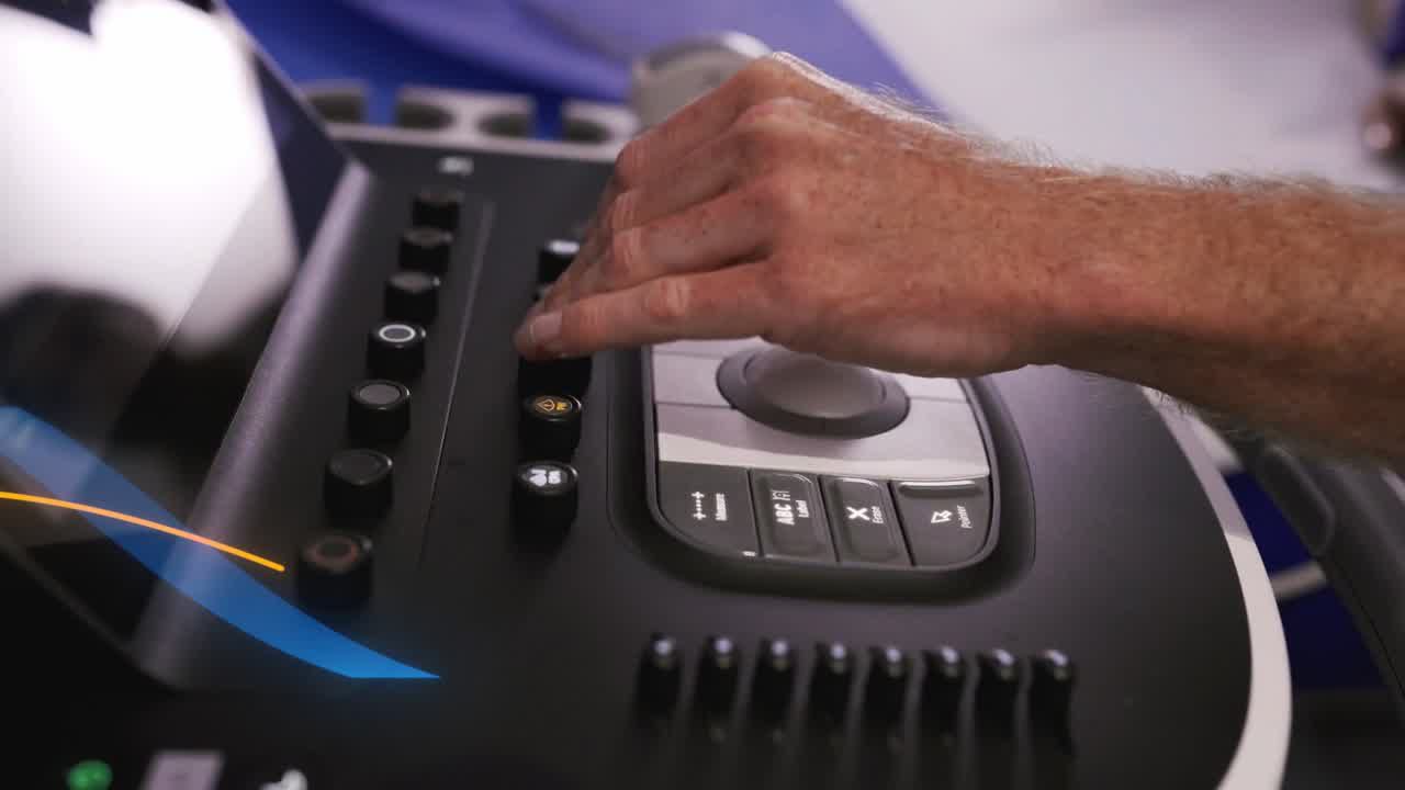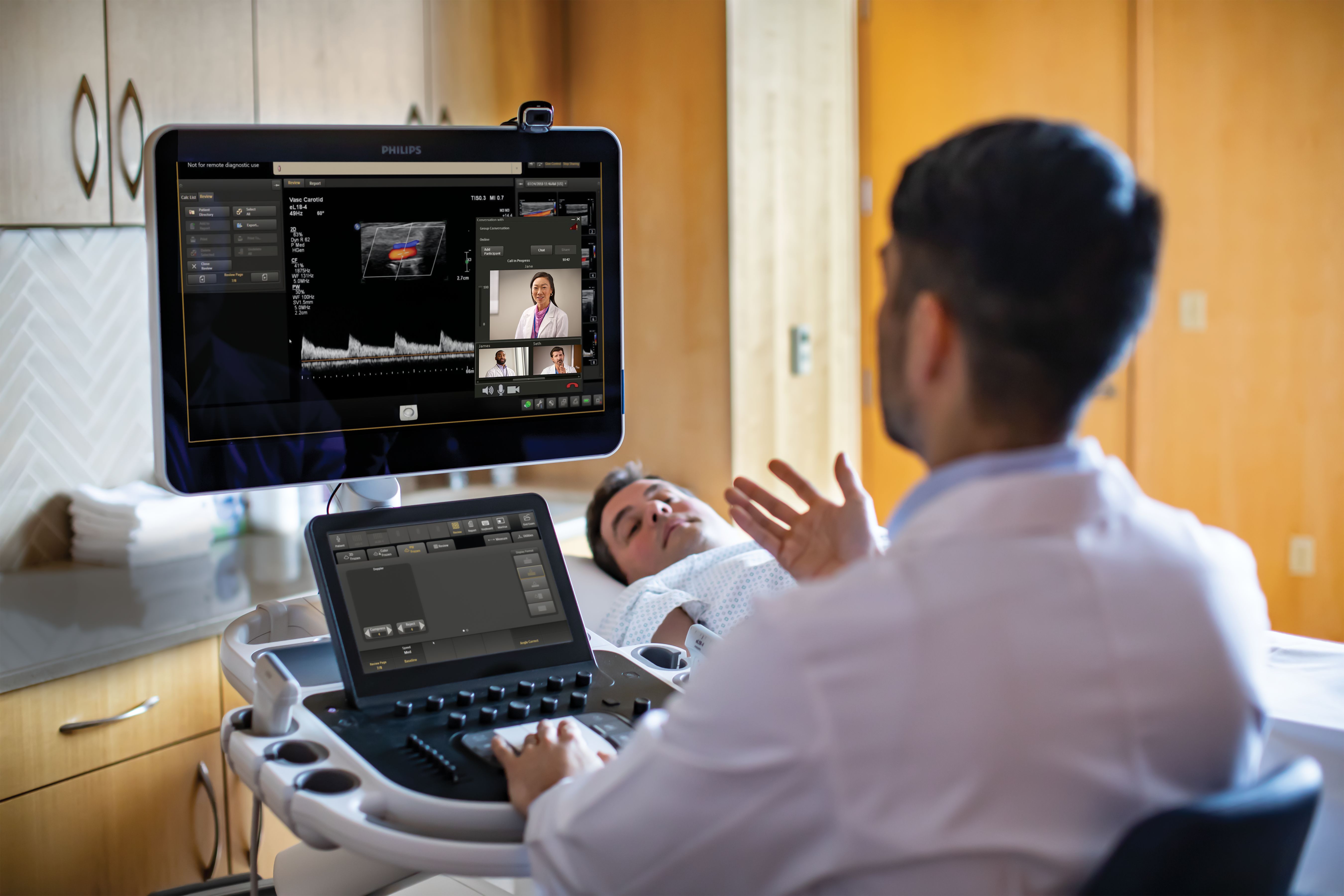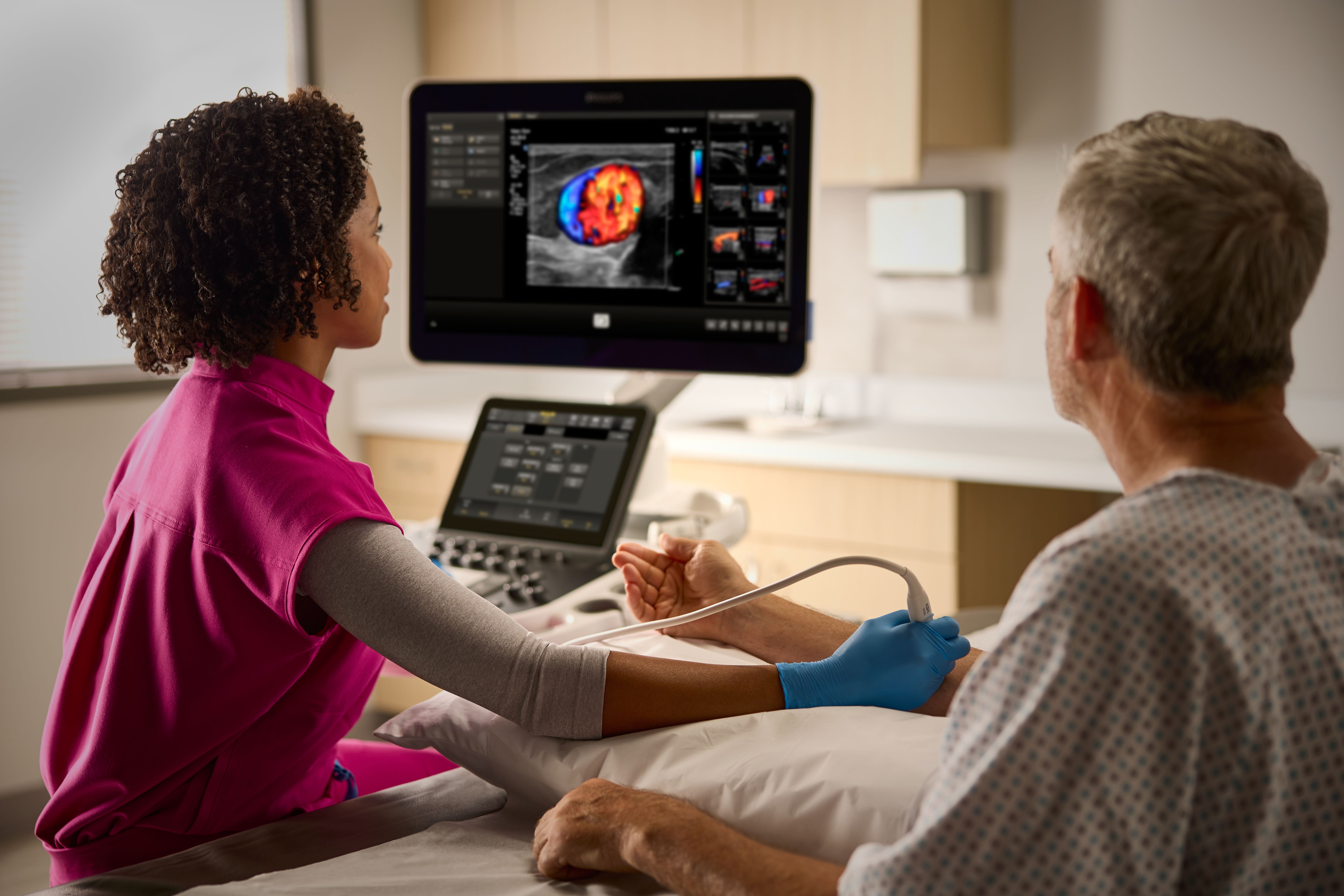
Hospitals and healthcare systems are continually being challenged to provide a higher quality of care cost-effectively. Premium ultrasound today demands improved clinical information from each scan, faster and more consistent exams that are easier to perform, and a higher level of confidence, even for technically difficult patients. The goal is quick and accurate diagnosis the first time and in less time. Philips EPIQ ultrasound features an exceptional level of clinical performance, workflow, and advanced intelligence to meet the challenges of today’s most demanding practices. The EPIQ platform brings ultimate solutions to ultrasound, with clinically tailored tools designed to elevate diagnostic confidence to new levels.
Many clinicians depend on ultrasound to elevate diagnostic confidence in detecting breast cancers in women. With mammography, dense breast tissue can mask small cancerous lesions and clinicians rely on ultrasound to enhance detection of lesions. The Philips ultimate ultrasound breast solution provides an all-in-one approach to empower clinicians to effectively assess, monitor and treat breast diseases, increasing diagnostic confidence and helping patients get the treatment they need.
Small Parts Imaging is one of the fastest growing segments of ultrasound exams for clinicians around the world. Its wide array of applications include assessing diseases and disorders of superficial organs like breasts, testicles and thyroid, as well as assessing musculoskeletal injuries like sprains and tears, making Small Parts ultrasound a vital tool for improving patient care. As a leader in health technology, Philips has developed the first ultrasound solution for Small Parts assessment, providing all-in-one functionality with both exceptional imaging and complementary clinical tools. Four key features work together to effectively assess, monitor and treat Small Parts, helping clinicians deliver exceptional patient care.
PureWave Imaging The Philips eL18-4 transducer incorporates both our highest frequency and ultra-broadband acoustic specification in a PureWave array design. This innovative transducer features a multi-row array configuration for full electronic focusing of the elevation plane. Elevation focusing works in conjunction with azimuthal focusing to provide thin-slice imaging for exceptional detail resolution and tissue uniformity from near to far depth of field. This supports a diverse range of clinical applications while delivering extraordinary imaging and depth-of-field performance. Full solution elastography The eL18-4 transducer is designed to support a complete elastography solution. Highly sensitive strain imaging can be used to rapidly assess relative tissue stiffness values across a variety of applications and shear wave elastography utilizes a unique pulsing scheme to generate and detect the propagation speed of shear waves, providing an absolute measure of tissue stiffness. The ability to combine both methods of elastography and deliver excellent imaging performance is an extraordinary clinical accomplishment that helps advance clinical practice. MicroFlow Imaging The eL18-4 supports Philips MicroFlow Imaging, a new proprietary method for blood flow detection providing an innovative approach to assess vascular beds. MicroFlow Imaging overcomes many of the barriers associated with conventional methods to detect small vessel blood flow with high resolution and minimal artifacts. MicroFlow Imaging maintains high frame rate and image quality and applies advanced artifact reduction techniques. 2D image subtraction, blending and side/side display options offer excellent visualization versatility across multiple clinical applications. Precision Biopsy Biopsy procedures, for many practices, are a routine part of an ultrasound examination. Using precision-guided biopsy techniques provides confidence in obtaining tissue targets to reduce multiple needle passes. The eL18-4 is compatible with the CIVCO Verza Guidance System, providing an advanced biopsy guidance system with virtually no dead zone. In addition, needle visualization software optimizes display of needle reflections for enhanced confidence during procedures.
It’s no secret that liver disease is a growing global health concern. According to the Centers for Disease Control and Prevention, liver disease is the 12th leading cause of death in the US. In fact, many people have hepatitis C and may not even know it. For decades, liver biopsy has been the conventional method for disease assessment, but the pain, expense and wait-time associated with results have proven to be barriers for some patients. Thanks to Philips innovations on the EPIQ ultrasound system, there is now a comprehensive all-in-one solution to help clinicians assess, treat and monitor many liver conditions.
PureWave Imaging PureWave crystal technology represents the biggest breakthrough in piezoelectric transducer material in 40 years. The pure, uniform crystals of PureWave are 85% more efficient than conventional piezoelectric material, resulting in exceptional performance. This technology is integral in our C5-1, C9-2 and X6-1 transducers allowing for improved penetration in difficult patients while maintaining excellent detail resolution, Doppler sensitivity and CEUS performance. ElastQ Imaging shear wave elastography Ultrasound shear wave elastography provides a noninvasive, reproducible, and easily performed method of assessing tissue stiffness. Research suggests that instead of a costly and painful biopsy procedure, an easy ultrasound exam including shear wave elastography could become routine for assessing liver disease status. ElastQ Imaging high-performance shear wave elastography features real-time, large Region of Interest (ROI) color-coded quantitative assessment of tissue stiffness. ElastQ Imaging also includes the ability to make retrospective measurements on stored images and a unique confidence map display uses intelligent analysis to add additional assurance that user measurements are obtained on tissue areas with adequate shear wave propagation. Contrast enhanced ultrasound Ultrasound contrast agents can transform the role of ultrasound in the liver, allowing the user to study the enhancement patterns of suspicious liver lesions in real time, providing access to faster and more confident diagnoses. With Philips EPIQ, CEUS is seamlessly integrated into the standard workflow, providing exceptional detail throughout arterial, portal, and late-phase scanning. Additionally, with advanced technologies at your fingertips – such as MaxVue FHD display, PureWave C9-2 transducer, fusion imaging, and Q-App quantification – EPIQ delivers maximum confidence for lesion detection and characterization, even in the most challenging exams. Image fusion and navigation Image fusion combines the inherent advantages of multimodality imaging directly on the ultrasound system using electromagnetic tracking. By combining CT/MR/PET with the live ultrasound and real-time position of the patient, the clinician has access to a powerful diagnostic tool while reducing radiation burden and maximizing throughput in the department. EPIQ’s exclusive Anatomical Intelligence Ultrasound (AIUS) comes to image fusion with automated registration of CT/MR and ultrasound volumes– in 1/10 the standard alignment time. Needle Navigation is a performance-enhancing tool for challenging interventional cases in the liver, and has been shown to improve workflow and reduce radiation exposure by using fewer confirmatory scans. Philips EPIQ with features a full suite of solutions which includes the NEW CIVCO eTrax™, Philips Adaptive Needle Tracker, and Philips Co-axial Needle Trackers. With such a wide range of compatibility to biopsy and ablation devices, the clinician has freedom to choose the best tools for the procedure depending on the level of complexity.
The filter criteria you have selected did not deliver any results
Please adjust your filters.
If you have any questions, please
contact us
You are about to visit a Philips global content page
Continue