Ultrasound AI Solutions
Achieve intuitive, reproducible ultrasound – scan to scan, user-to user
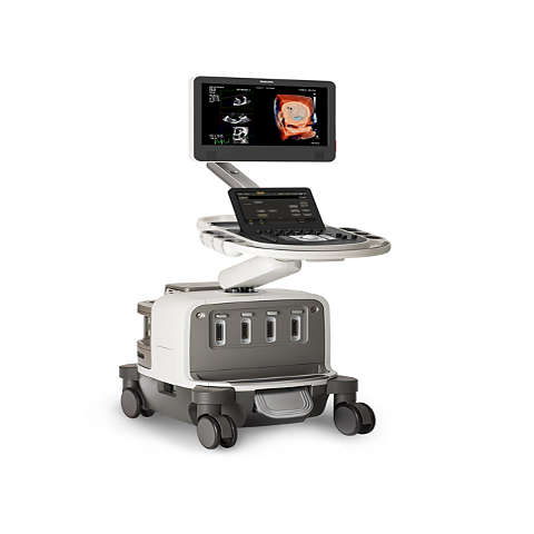
Exceptional images, quantification and automation
The EPIQ CVx offers advanced imaging and quantification and customizable capabilities that can significantly reduce exam time and speed time-to-results, including for transthoracic (TTE) or transesophageal (TEE) echo, so that you can provide greater care in less time for more types of patients.
Image gallery
- Toggle view
Features

Automatic color flow quantification to assess mitral regurgitation
3D Auto Color Flow Quantification uses AI for fast, easy and reproducible mitral regurgitation (MR) volume to help assess MR severity. It has been proven to work on single, multiple, concentric and eccentric jets.

AI-driven tricuspid valve quantification to guide device selection
3D Auto Tricuspid Valve Quantification uses AI to help evaluate annulus size to guide device selection. It facilitates more accurate peri-procedure TV annulus measurements and helps you leverage initial sizing and plan with CT.
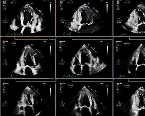
AI-based software for the preselection and analysis of optimal apical cardiac ultrasound views
SmartView Select AI-based technology is designed to run behind the scenes while a clinician is capturing images. It seeks and identifies the effective images to use in certain applications such as Strain or EF, as needed by the user. This helps reduce the variability associated with manual view selection and visual analysis of cardiac ultrasound images.
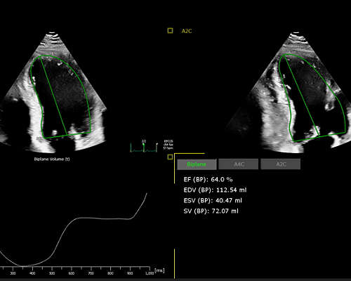
AI-powered automated ejection fraction (EF) calculation for objective EF analysis
2D Auto EF* AI-based technology is designed to reduce variability in the calculation of EF from 4CH and/or 2CH views and Biplane and improve efficiency through automation. It allows users to assess cardiac function by any clinician, with all levels of experience with qualitative assessment.
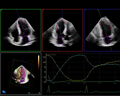
Cardiac 3D chamber quantifications driven by advanced automation
Dynamic HeartModel** tracks every frame over the cardiac cycle using 3D speckle technology to provide a holistic view of the left heart function. Automated border detection, multi-beat selection and results average yield a more reliable heart function evaluation than single beat in arrhythmia patients.

Comprehensive mitral valve modeling—live 3D volume measurements and calculations
The Mitral Valve Navigator*** is designed to take a Live 3D volume of the mitral valve and turn it into an easy-to-interpret model in eight guided steps, providing access to a comprehensive list of MV measurements and calculations. Internal comparison of MVQ to MVNA tools measures 89% fewer clicks.1
Automation for robust, proven reproducible cardiac quantification in both 2D and 3D
AutoStrain delivers fast reproducible 2D strain quantification for the LV, LA and RV. Philips AutoMeasure provides fully automated 2D (length and EF and Doppler measurements. Philips 3D Auto RV offers full 3D quantification for RV volumes and functional assessment.
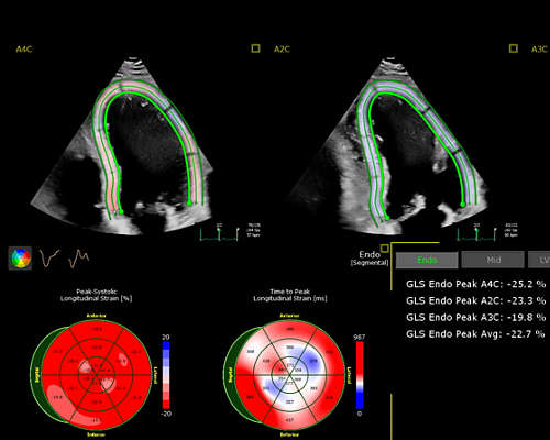

AI-based border detection and score calculation for LV impairment
Segmental Wall Motion Scoring is an AI-based software that automatically detects borders and uses all left ventricular (LV) segments to calculate a score that aids assessment of LV impairment.
Ultrasound AI Solutions technology is available on
-
EPIQ CVx
EPIQ CVx, our premium cardiovascular ultrasound system built on our innovative, modular, industry-leading ultrasound platform, has powerful AI-based capabilities and advanced diagnostic solutions to help you transcend today's complexities and propel echocardiography into the next dimension. This enables you to achieve greater consistency, accessible innovation, smarter workflows, and easier scalability.
795231 -
Affiniti CVx
Affiniti CVx, built on the Philips innovative cardiovascular ultrasound platform, has powerful AI-based capabilities to help you transcend today's complexities and propel echocardiography into the next dimension. Affiniti CVx offers smart features to enable you to achieve greater consistency, accessible innovation, smarter workflows and easier scalability. This is all on one familiar, industry-leading platform so you can act and decide with the ease you know and the legacy you trust.
795190
Documentation
- Resources
-
Related stories
Footnotes
*2D Auto EF from DiA Imaging analysis, a Philips company, is a feature of Philips CV Ultrasound **Dynamic HeartModel is an advanced automation solution with anatomical intelligence, not currently certified as artificial intelligence. *** Mitral Valve Navigator is an advanced automation solution with anatomical intelligence, not currently certified as artificial intelligence. 1Henry MP, et al., Three-Dimensional Transthoracic Static and Dynamic Normative Values of the Mitral Valve Apparatus: Results from the Multicenter World Alliance Societies of Echocardiography Study. J Am Soc Echocardiogr. 2022 Jul;35(7):738-751.e1. doi: 10.1016/j.echo.2022.02.010. Epub 2022 Mar 1. PMID: 35245668; PMCID: PMC10257802.
